
'/%3E%3C/svg%3E)

'/%3E%3Cpath%20fill='%23b2b2b2'%20d='M241%20382q0%2016%2017%2026%2017%2011%2042%2011t42-11q16-11%2017-26V269H241'/%3E%3Cpath%20fill='%239f7e00'%20d='M241%20379q0%2014%208%2023%209%209%2021%209%2014%200%2021-9%209-9%209-23V269h-59'/%3E%3Cpath%20d='M241%20339v-70h59v110q0%2014-9%2023l-6%205v-68h-15v72q-8%200-14-4v-68zm59%2040c0%2022%2015%2032%2029%2032%2021%200%2030-18%2030-32v-40h-59'/%3E%3Cpath%20fill='%239f7e00'%20d='M266%20277v5h2v12h-3v-5h2v-5h-9v5h2v5h-4v9h3v18h-5v10h34v-10h-5v-18h3v-9h-4v-5h2v-5h-9v5h2v5h-3v-12h2v-5zm42%20101q0%2010%206%2017%206%206%2015%207%2010-1%2016-7%206-7%206-17v-29h-43'/%3E%3Ccircle%20fill='%230044ad'%20cx='329.5'%20cy='372'%20r='6'/%3E%3Cellipse%20fill='%230044ad'%20cx='300'%20cy='340'%20rx='16'%20ry='21'/%3E%3C/svg%3E)


'/%3E%3C/g%3E%3Cuse%20href='%23c'%20transform='matrix(-1%200%200%201%20720%200)'/%3E%3C/svg%3E)





Traumatic Diabetic Wound with NPWT and Polyhexanide + Poloxamer
A documented traumatic diabetic foot wound case involving necrotic tissue debridement, Polyhexanide + Poloxamer solution, NPWT support, full-thickness skin grafting and Polyhexanide + Poloxamer gel-based dressing follow-up.
Essential Case Information
This overview summarizes the case in a structured format: who, what, where, when, why and how the traumatic diabetic wound care pathway was managed.
The Patient
A 47-year-old male patient with known hypertension and diabetes mellitus history.
The Clinical Problem
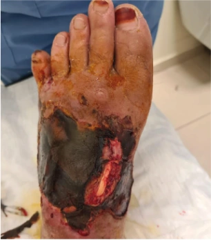
A traumatic wound on the dorsum of the foot after falling from height, measuring approximately 10 × 12 cm with necrotic tissue and preserved paratenon.
The Setting
The case was documented by the Plastic, Reconstructive and Aesthetic Surgery Department, Wound Healing Unit, Kahramanmaraş Sütçü İmam University Faculty of Medicine.
The Timeline
After debridement and wound preparation, NPWT was applied in 3-day intervals. Granulation was observed by day 15, followed by grafting and epithelialization.
The Objective
The objective was to support traumatic diabetic wound management through debridement, infection assessment, Polyhexanide + Poloxamer cleansing and NPWT-assisted wound bed preparation.
The Care Pathway
Necrotic tissue was debrided, infection parameters and tissue culture were evaluated, the wound was treated with Actolind® Solution Plus and NPWT was applied before grafting.
Objective
This case presents the combined use of Polyhexanide + Poloxamer-based wound cleansing and Negative Pressure Wound Therapy in a traumatic diabetic foot wound. The aim was to support wound bed preparation, infection-risk management, granulation and epithelialization through a structured wound care plan.
Case Summary
A 47-year-old male patient with hypertension and diabetes mellitus presented with a traumatic wound on the dorsum of the foot after falling from height. The wound was approximately 10 × 12 cm, necrotic on the surface and had preserved paratenon.
Necrotic tissue debridement was performed. Hemogram, biochemistry, sedimentation and CRP were evaluated as infection parameters. Deep tissue culture was taken, and no growth was observed. Infection parameters were reported within normal limits.
Wound Care Method
- Necrotic tissue was debrided.
- Hemogram, biochemistry, sedimentation and CRP were evaluated.
- Deep tissue culture was obtained.
- No growth was observed in the wound culture.
- The wound was treated with Actolind® Solution Plus.
- The solution was left in contact with the wound for 5 minutes.
- NPWT was applied every 3 days.
- After granulation, full-thickness skin grafting was performed.
- After graft dressing removal, epithelialization was observed.
- Actolind® Gel Plus was recommended for daily dressing after discharge.
Clinical Follow-up
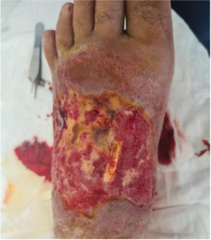
The wound was followed with NPWT in 3-day intervals after Polyhexanide + Poloxamer cleansing. By the 15th day, the wound bed was reported as granulated. A full-thickness skin graft was then applied and kept dressed for 5 days.
Outcome
After the dressing was opened, epithelialization of the wound was observed. The patient was discharged with recommendations for daily dressing using Actolind® Gel Plus.
Traumatic Diabetic Wound Image Areas
Use these image fields for the original case visuals. Image paths and labels can be changed from the settings area at the top of this HTML block.
06.06.2022
 12.06.2022
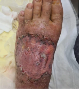 26.06.2022
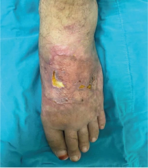 07.10.2022
Documented Follow-up Steps
The timeline below summarizes the reported wound care process and key clinical milestones.
Traumatic Wound Assessment
A 10 × 12 cm necrotic traumatic wound on the dorsum of the foot was evaluated in a diabetic patient.
Necrotic Tissue Removal
Necrotic tissue was debrided, and infection parameters with deep tissue culture were evaluated.
No Growth Reported
Deep tissue culture showed no growth, and infection parameters were reported as normal.
NPWT Follow-up
Actolind® Solution Plus was applied with 5-minute contact time, followed by NPWT every 3 days.
Granulation Observed
By the 15th day, granulation of the wound bed was observed.
Full-Thickness Skin Graft
A full-thickness skin graft was applied and kept dressed for 5 days.
Daily Gel Dressing Recommendation
After epithelialization was observed, the patient was discharged with Actolind® Gel Plus daily dressing recommendations.
Outcome Highlights
Explore More Actolind® Clinical Case Resources
Review additional wound care case materials, product information and professional resources prepared for healthcare professionals.
View All Case Studies