
'/%3E%3C/svg%3E)

'/%3E%3Cpath%20fill='%23b2b2b2'%20d='M241%20382q0%2016%2017%2026%2017%2011%2042%2011t42-11q16-11%2017-26V269H241'/%3E%3Cpath%20fill='%239f7e00'%20d='M241%20379q0%2014%208%2023%209%209%2021%209%2014%200%2021-9%209-9%209-23V269h-59'/%3E%3Cpath%20d='M241%20339v-70h59v110q0%2014-9%2023l-6%205v-68h-15v72q-8%200-14-4v-68zm59%2040c0%2022%2015%2032%2029%2032%2021%200%2030-18%2030-32v-40h-59'/%3E%3Cpath%20fill='%239f7e00'%20d='M266%20277v5h2v12h-3v-5h2v-5h-9v5h2v5h-4v9h3v18h-5v10h34v-10h-5v-18h3v-9h-4v-5h2v-5h-9v5h2v5h-3v-12h2v-5zm42%20101q0%2010%206%2017%206%206%2015%207%2010-1%2016-7%206-7%206-17v-29h-43'/%3E%3Ccircle%20fill='%230044ad'%20cx='329.5'%20cy='372'%20r='6'/%3E%3Cellipse%20fill='%230044ad'%20cx='300'%20cy='340'%20rx='16'%20ry='21'/%3E%3C/svg%3E)


'/%3E%3C/g%3E%3Cuse%20href='%23c'%20transform='matrix(-1%200%200%201%20720%200)'/%3E%3C/svg%3E)





Diabetic Foot Wound Care
A diabetic foot wound care case resource involving systematic wound cleansing with Actolind® w Solution, topical support with Actolind® w Gel, regular dressing follow-up and photographic wound monitoring.
Essential Case Information
This overview summarizes the case in a structured format: who, what, where, when, why and how the diabetic foot wound care pathway was managed.
The Patient
A diabetic patient with a chronic foot or lower extremity wound requiring structured wound care, cleansing and regular follow-up.
The Clinical Problem
A diabetic foot wound with impaired wound bed condition, exudate or tissue burden, requiring systematic cleansing, topical support and dressing management.
The Setting
The case belongs to the diabetic foot and lower extremity wound care documentation within Actolind® clinical case materials.
The Timeline
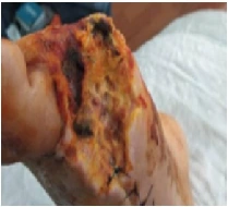
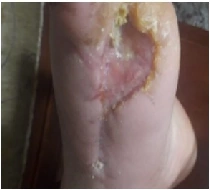
Wound progress was followed through serial clinical photographs and dressing changes adjusted according to wound status, exudate level and clinical need.
The Objective
The objective was to support practical diabetic foot wound management through regular cleansing, topical wound care and professional follow-up.
The Care Pathway
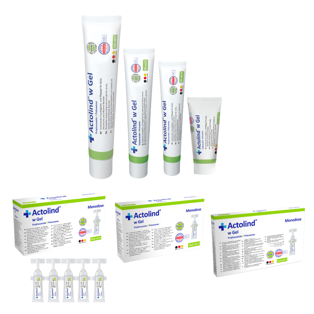
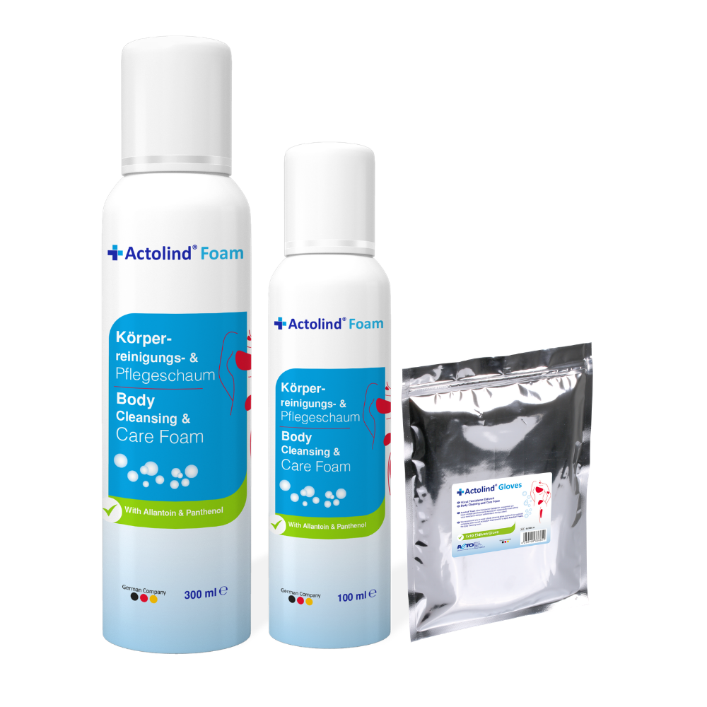
The wound was cleansed with Actolind® w Solution, supported with Actolind® w Gel and covered with appropriate primary and secondary dressings.
Objective
This case resource presents the use of Actolind® w Solution and Actolind® w Gel as part of professional diabetic foot wound care. The objective is to demonstrate a practical care pathway involving wound cleansing, topical support, dressing follow-up and photographic documentation.
Case Summary
Diabetic foot wounds are commonly associated with delayed wound healing, infection risk, impaired tissue perfusion, neuropathy and pressure-related trauma. In the documented case materials, diabetic foot and lower extremity wounds were followed with regular cleansing, topical gel application and appropriate dressing selection.
Wound care was adapted according to the wound bed condition, exudate level and clinical progression. Serial photographic follow-up was used to document the wound course and visual changes over time.
Wound Care Method
- The wound was assessed before each dressing change.
- Wound cleansing or irrigation was performed with Actolind® w Solution.
- After cleansing, Actolind® w Gel was applied to the wound bed.
- A suitable non-adherent primary dressing was used when required.
- Secondary dressing was applied according to exudate and wound condition.
- Dressing frequency was adjusted according to the wound status.
- Photographic follow-up was used to document wound progression.
Clinical Follow-up
During the follow-up period, the wound bed appearance, exudate level, tissue quality and progression of epithelialization were monitored. Dressing intervals were adjusted according to clinical need, and the wound care approach was continued as part of routine professional wound management.
Outcome
The documented diabetic foot and lower extremity wound cases showed visible improvement in wound bed appearance over time. The results should be interpreted together with patient-specific factors such as diabetes duration, glycemic control, infection risk, vascular status, pressure management and standard clinical care.
Diabetic Foot Wound Image Areas
Use these image fields for the original case visuals. Image paths and labels can be changed from the settings area at the top of this HTML block.
Clinical View 1
 Clinical View 2
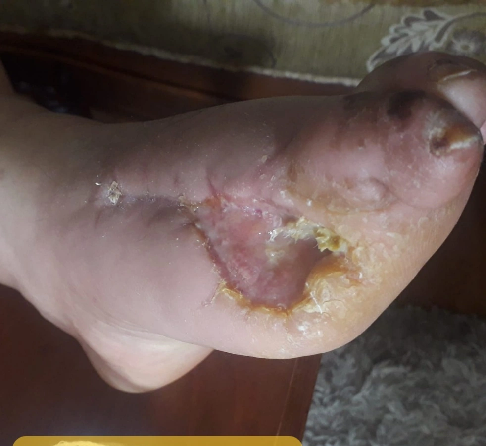 Clinical View 3
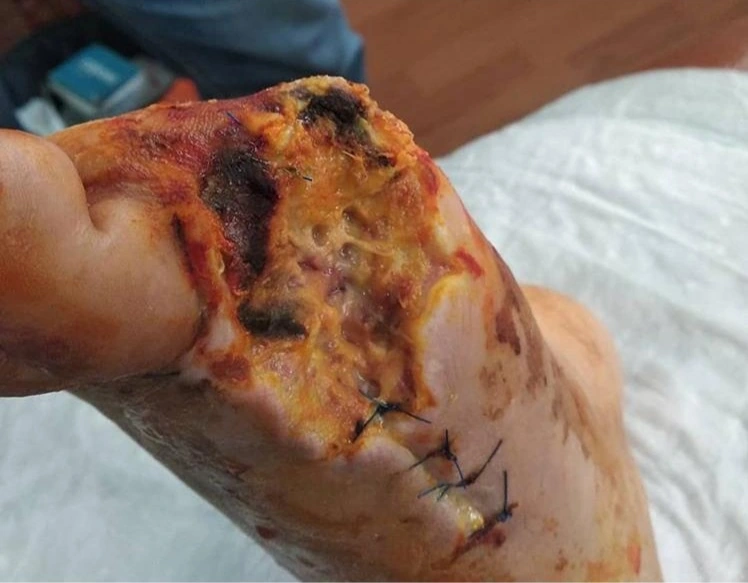 Clinical View 4
Documented Follow-up Steps
The timeline below summarizes the practical diabetic foot wound care process.
Initial Wound Assessment
The wound bed condition, exudate level, surrounding skin and patient-specific risk factors were evaluated before starting the dressing plan.
Systematic Cleansing
The wound was cleansed or irrigated with Actolind® w Solution during dressing changes.
Topical Gel Application
Actolind® w Gel was applied to the wound bed after cleansing as topical wound care support.
Dressing and Protection
A suitable non-adherent primary dressing and secondary dressing were used according to wound condition and exudate level.
Photographic Follow-up
Serial photographs were used to document wound bed changes, tissue progression and practical wound management outcomes.
Outcome Highlights
Explore More Actolind® Clinical Case Resources
Review additional wound care case materials, product information and professional resources prepared for healthcare professionals.
View All Case Studies