
'/%3E%3C/svg%3E)

'/%3E%3Cpath%20fill='%23b2b2b2'%20d='M241%20382q0%2016%2017%2026%2017%2011%2042%2011t42-11q16-11%2017-26V269H241'/%3E%3Cpath%20fill='%239f7e00'%20d='M241%20379q0%2014%208%2023%209%209%2021%209%2014%200%2021-9%209-9%209-23V269h-59'/%3E%3Cpath%20d='M241%20339v-70h59v110q0%2014-9%2023l-6%205v-68h-15v72q-8%200-14-4v-68zm59%2040c0%2022%2015%2032%2029%2032%2021%200%2030-18%2030-32v-40h-59'/%3E%3Cpath%20fill='%239f7e00'%20d='M266%20277v5h2v12h-3v-5h2v-5h-9v5h2v5h-4v9h3v18h-5v10h34v-10h-5v-18h3v-9h-4v-5h2v-5h-9v5h2v5h-3v-12h2v-5zm42%20101q0%2010%206%2017%206%206%2015%207%2010-1%2016-7%206-7%206-17v-29h-43'/%3E%3Ccircle%20fill='%230044ad'%20cx='329.5'%20cy='372'%20r='6'/%3E%3Cellipse%20fill='%230044ad'%20cx='300'%20cy='340'%20rx='16'%20ry='21'/%3E%3C/svg%3E)


'/%3E%3C/g%3E%3Cuse%20href='%23c'%20transform='matrix(-1%200%200%201%20720%200)'/%3E%3C/svg%3E)





Diabetic Foot Postoperative Wound with PRF
A documented postoperative diabetic foot wound case involving infection follow-up, Polyhexanide + Poloxamer solution and gel, PRF dressing support and complete epithelialization by day 14.
Essential Case Information
This overview summarizes the case in a structured format: who, what, where, when, why and how the postoperative diabetic foot wound care pathway was managed.
The Patient
A 57-year-old male patient with known diabetes mellitus and hypertension history.
The Clinical Problem
A wound opening after flap surgery in a diabetic foot case, with elevated CRP and positive wound culture findings.
The Setting
The case was documented by the Plastic, Reconstructive and Aesthetic Surgery Department, Wound Healing Unit, Kahramanmaraş Sütçü İmam University Faculty of Medicine.
The Timeline
Initial infection assessment was followed by daily solution and gel care. After 5 days, CRP normalized and wound culture showed no growth. PRF dressing was then continued every 3 days, and epithelialization was observed by day 14.
The Objective
The objective was to control infection risk, support the postoperative wound bed and promote epithelialization using Polyhexanide + Poloxamer products and PRF.
The Care Pathway
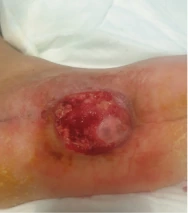
The wound was treated with gauze soaked in Actolind® w Solution for 5 minutes, followed by daily Actolind® w Gel dressing and PRF dressing every 3 days after infection control.
Objective
This case presents a postoperative diabetic foot wound managed with Polyhexanide + Poloxamer solution and gel, infection follow-up and PRF dressing support. The aim was to support infection control, wound bed improvement and epithelialization after flap surgery.
Case Summary
A 57-year-old male patient with diabetes mellitus and hypertension was referred with a wound opening at the flap site after flap surgery. Infection parameters including CRP, sedimentation and hemogram were evaluated. The CRP value was reported as 75, and the patient was consulted with the infection department.
Empirical treatment was started until culture results were available. Wound culture showed Gram-negative bacilli, with Pseudomonas aeruginosa and Acinetobacter baumannii / calcoaceticus complex reported. Antibiotic therapy was then arranged according to the culture antibiogram.
Wound Care Method
- Infection parameters were evaluated, including CRP, sedimentation and hemogram.
- The patient was consulted with the infection department.
- Empirical treatment was started until culture results were available.
- Culture antibiogram-guided antibiotic therapy was initiated.
- The wound was treated with gauze soaked in Actolind® w Solution for 5 minutes.
- Daily dressing was performed with Actolind® w Gel.
- After infection parameters improved, PRF dressing was added.
- PRF dressing was continued every 3 days.
- Wound epithelialization was monitored during follow-up.
Clinical Follow-up
Five days after the initial wound care and infection management, the patient’s CRP value returned to normal and wound culture showed no growth. After this point, dressing care continued with PRF obtained from the patient’s blood at 3-day intervals.
Outcome
According to the original case documentation, complete epithelialization of the wound was observed by the 14th day. The case highlights the importance of infection control and structured postoperative wound care in diabetic patients.
Diabetic Foot PRF Image Areas
Use these image fields for the original case visuals. Image paths and labels can be changed from the settings area at the top of this HTML block.
06.07.2022
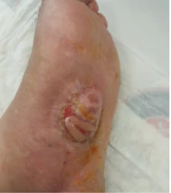 11.07.2022
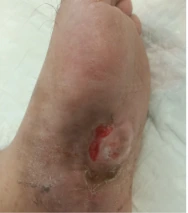 17.07.2022
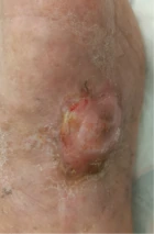 21.07.2022
Documented Follow-up Steps
The timeline below summarizes the reported wound care process and key clinical milestones.
Post-flap Wound Opening
The patient presented with wound opening after flap surgery. Infection parameters were evaluated.
Infection Consultation
CRP was elevated, and the patient was consulted with the infection department. Empirical treatment was started until culture results were available.
Microbiological Findings
Wound culture showed Gram-negative bacilli, including Pseudomonas aeruginosa and Acinetobacter baumannii / calcoaceticus complex.
Solution Contact Time
Gauze soaked with Actolind® w Solution was applied to the wound for 5 minutes.
Gel Dressing Care
Daily dressing was performed with Actolind® w Gel during the initial wound care period.
Infection Control Follow-up
CRP returned to normal and wound culture showed no growth after 5 days.
PRF Dressing Stage
PRF dressing obtained from the patient’s blood was continued every 3 days.
Complete Epithelialization
Complete epithelialization of the wound was observed by the 14th day.
Outcome Highlights
Explore More Actolind® Clinical Case Resources
Review additional wound care case materials, product information and professional resources prepared for healthcare professionals.
View All Case Studies