





Chronic Wound with A. baumannii Colonization
A documented case involving a ten-year chronic wound with A. baumannii growth, managed with contact isolation, asepsis and antisepsis principles, PHMB-based wound care, debridement and skin grafting.
Essential Case Information
This overview summarizes the case in a structured format: who, what, where, when, why and how the wound care pathway was managed.
The Patient
A patient with a ten-year history of chronic ulceration on the anterior surface of the left lower leg.
The Clinical Problem
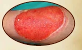
A chronic wound measuring 17 × 28 cm with regional erythema, dirty granulation tissue, indurated surrounding tissue and irregular wound borders.
The Setting
The patient was managed at Bağcılar Training and Research Hospital, Plastic, Reconstructive and Aesthetic Surgery Department.
The Timeline
Initial presentation was documented on 08 August 2008. Control cultures were taken during follow-up, and debridement with skin grafting was performed on 27 August 2008.
The Objective
The objective was to manage a difficult chronic wound with A. baumannii growth using isolation measures, wound care, nursing follow-up and surgical reconstruction.
The Care Pathway
Contact isolation, patient-specific dressing materials, infection-control nursing care, PHMB solution and gel, debridement and skin grafting were applied.
Objective
This case presents the management of a patient with a ten-year chronic wound in which A. baumannii was detected. The case demonstrates a combined approach involving appropriate isolation techniques, asepsis and antisepsis principles, nursing care, PHMB-based dressing procedures and surgical treatment.
Case Summary
The patient presented with a chronic ulcer on the anterior surface of the left lower leg. The wound measured 17 × 28 cm at admission. The admission culture showed A. baumannii growth. The patient was admitted to the Plastic, Reconstructive and Aesthetic Surgery service and placed in a single room under contact isolation.
All dressing materials were reserved for the patient. Wound care was performed for 10 days by infection control nurses in accordance with asepsis and antisepsis techniques. A 0.1% polyhexanide-containing solution and gel were used during dressing procedures.
Wound Care Method
- Admission wound culture was obtained.
- Contact isolation was implemented after A. baumannii detection.
- The patient was placed in a single room.
- Patient-specific dressing materials were used.
- Wound care was performed according to asepsis and antisepsis principles.
- 0.1% polyhexanide-containing solution and gel were used in dressing procedures.
- Wound dimensions and clinical progress were monitored during follow-up.
- Debridement followed by skin grafting was performed on 27 August 2008.
Findings
The wound was described as an open chronic wound with regional erythema, dirty granulation tissue, indurated surrounding tissue and irregular borders. Control culture taken on 18 August 2008 showed no growth. The last two cultures taken on 22–23 August 2008 also showed no growth.
Outcome
The wound size decreased from 17 × 28 cm to 13 × 20 cm during follow-up. Skin grafting was performed on 27 August 2008, and the graft was reported as 100% successful. No problems were reported in early or late follow-up.
10-Year Chronic Wound Image Series
Photographs extracted from clinical documentation during pre-graft care, wound progression, and post-graft evaluation.
Pre-graft View 1
 Pre-graft View 2
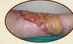 Care Progress 1
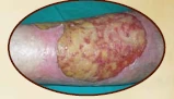 Care Progress 2
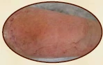 Post-graft View 1
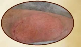 Post-graft View 2
Documented Follow-up Steps
The timeline below summarizes key clinical milestones reported in the original case material.
Initial Presentation and Culture
The patient presented with a 17 × 28 cm chronic wound. Admission culture was obtained, and A. baumannii was detected.
Structured Wound Care Period
Dressing procedures were performed by infection control nurses using asepsis and antisepsis principles, with PHMB solution and gel.
Control Culture
The control culture taken during follow-up showed no growth.
Final Follow-up Cultures
The last two cultures taken during follow-up also showed no growth.
Debridement and Skin Graft
Debridement was followed by skin grafting. The graft was reported as fully successful.
Outcome Highlights
Explore More Actolind® Clinical Case Resources
Review additional wound care case materials, product information and professional resources prepared for healthcare professionals.
View All Case Studies